A possible approach to early non-invasive diagnosis of ovarian cancer
Ovarian cancer (OC) has one of the highest incidence rates within the oncological pathology of the female reproductive system worldwide. Despite the wide range of diagnostic techniques, this pathology in most cases is diagnosed at the last stages which is associated with a poor prognosis and low survival of patients.
This paper is assigned to a detailed study of the morphology and the composition of specific ovarian cancer calcifications, which are composed of hydroxyapatite. We also provide a comparative analysis of calcification sizes and the resolution of minimally invasive diagnostic methods. Since nanosized calcifications appear in tumor tissue at early stages, it could be a basis for the development of new technology for screening and early diagnostics of ovarian cancer. Therefore, the study of hydroxyapatite nanocrystallite's properties will reveal the best method for early ovarian cancer diagnostics.
Calcification or biomineralization is one of the clinical and morphological features of ovarian tumor manifestation. Pathological biomineralization (PBM) is detected in about 8% of OC cases by computer tomography. Calcification is more common for serous ovarian adenocarcinoma. Histologically, the incidence of calcifications in low-grade and high-grade serous carcinomas is 100% and 50%, respectively. Pathological biomineral deposits start to develop in the earliest stages of carcinogenesis as nanocrystalline objects.
The study of the microstructure and phase composition of these crystallites is required for a deeper understanding of the processes of calcification in ovarian tumors. It may be the basis for the introduction of new early diagnosis and/or treatment methods.
We compared the results of ultrasound and histology and found that calcifications of a size less than 200 μm are not detected by ultrasound. At the same time, CT (computed tomography) scans detect pathological deposits of 1000 μm and bigger. IVUS (intravascular ultrasound) visualizes calcification with a size of more than 100 μm. OCT (optical coherence tomography) can recognize biomineral inclusions with a size of 10–20 μm and more. Technological advances make it possible to diagnose ever-smaller objects. This obviously depends on the composition of pathological biominerals, as well as on the maturity of the biomineral and its structure.
The OC calcified structures are round fragile particles of different sizes. In the EDX (Energy-dispersive X-ray spectroscopy) spectra, the main lines were from Ca and P, and the ratio of these elements corresponds to hydroxyapatite. According to TEM, pathological crystalline nanoparticles are polydisperse and their size ranges from 5 to 50 nm.
Also we found that tumors without PBM had significantly bigger sizes. This indicates a possible positive role (tumor suppression) of calcifications in ovarian tumors. There is a strong correlation between tumor size and calcification. Thus, the detection of nanocrystalline particles will contribute to the detection of tumors at the initial stages of cancerogenesis. This confirms our hypothesis about the possible application of high-resolution techniques for early cancer diagnostics.
Therefore, the study of the structure and physical, chemical and phase composition of OC calcifications, as well as features of their visualization, is important since provides the possible practical application of this pathological phenomenon for early diagnostics of OC and other neoplasms with biomineralization.
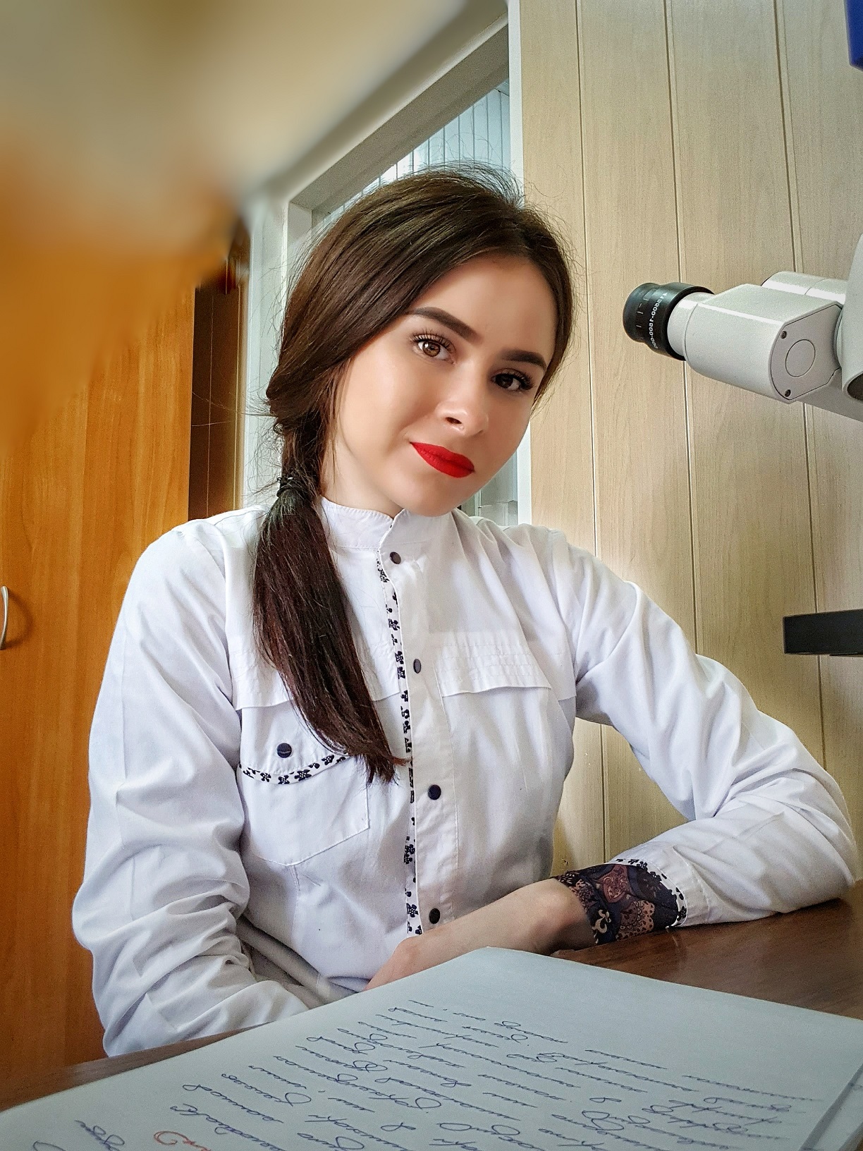
-Ruslana Chyzhma, Medical Institute of Sumy State University, Pathology Department Resident
Full text:
Potential Role of Hydroxyapatite Nanocrystalline for Early Diagnostics of Ovarian Cancer - https://www.mdpi.com/2075-4418/11/10/1741/htm

